Friday, 19 June 2020

Diluting blood plasma rejuvenates tissue, reverses aging in mice


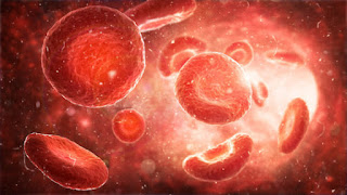
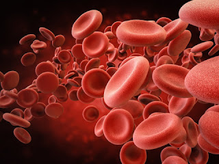
Red blood cells in artery illustration

In 2005, University of California, Berkeley, researchers made the surprising discovery that making conjoined twins out of young and old mice -- such that they share blood and organs -- can rejuvenate tissues and reverse the signs of aging in the old mice. The finding sparked a flurry of research into whether a youngster's blood might contain special proteins or molecules that could serve as a "fountain of youth" for mice and humans alike.

But a new study by the same team shows that similar age-reversing effects can be achieved by simply diluting the blood plasma of old mice -- no young blood needed.

In the study, the team found that replacing half of the blood plasma of old mice with a mixture of saline and albumin -- where the albumin simply replaces protein that was lost when the original blood plasma was removed -- has the same or stronger rejuvenation effects on the brain, liver and muscle than pairing with young mice or young blood exchange. Performing the same procedure on young mice had no detrimental effects on their health.

This discovery shifts the dominant model of rejuvenation away from young blood and toward the benefits of removing age-elevated, and potentially harmful, factors in old blood.

"There are two main interpretations of our original experiments: The first is that, in the mouse joining experiments, rejuvenation was due to young blood and young proteins or factors that become diminished with aging, but an equally possible alternative is that, with age, you have an elevation of certain proteins in the blood that become detrimental, and these were removed or neutralized by the young partners," said Irina Conboy, a professor of bioengineering at UC Berkeley who is the first author of the 2005 mouse-joining paper and senior author of the new study. "As our science shows, the second interpretation turns out to be correct. Young blood or factors are not needed for the rejuvenating effect; dilution of old blood is sufficient."

In humans, the composition of blood plasma can be altered in a clinical procedure called therapeutic plasma exchange, or plasmapheresis, which is currently FDA-approved in the U.S. for treating a variety of autoimmune diseases. The research team is currently finalizing clinical trials to determine if a modified plasma exchange in humans could be used to improve the overall health of older people and to treat age-associated diseases that include muscle wasting, neuro-degeneration, Type 2 diabetes and immune deregulation.

"I think it will take some time for people to really give up the idea that that young plasma contains rejuvenation molecules, or silver bullets, for aging," said Dobri Kiprov, a medical director of Apheresis Care Group and a co-author of the paper. "I hope our results open the door for further research into using plasma exchange -- not just for aging, but also for immunomodulation."

The study appears online in the journal Aging.

A molecular 'reset' button

In the early 2000s, Conboy and her husband and research partner Michael Conboy, a senior researcher and lecturer in the Department of Bioengineering at UC Berkeley and co-author of the new study, had a hunch that our body's ability to regenerate damaged tissue remains with us into old age in the form of stem cells, but that somehow these cells get turned off through changes in our biochemistry as we age.

"We had the idea that aging might be really more dynamic than people think," Conboy said. "We thought that it could be caused by transient and very reversible declines in regeneration, such that, even if somebody is very old, the capacity to build new tissues in organs could be restored to young levels by basically replacing the broken cells and tissues with healthy ones, and that this capacity is regulated through specific chemicals which change with age in ways that become counterproductive."

After the Conboys published their groundbreaking 2005 work, showing that making conjoined twins from the old mouse and a young mouse reversed many signs of aging in the older mouse, many researchers seized on the idea that specific proteins in young blood could be the key to unlocking the body's latent regeneration abilities.

However, in the original report, and in a more recent study, when blood was exchanged between young and old animals without physically joining them, young animals showed signs of aging. These results indicated that that young blood circulating through young veins could not compete with old blood.

As a result, the Conboys pursued the idea that a buildup of certain proteins with age is the main inhibitor of tissue maintenance and repair, and that diluting these proteins with blood exchange could also be the mechanism behind the original results. If true, this would suggest an alternative, safer path to successful clinical intervention: Instead of adding proteins from young blood, which could do harm to a patient, the dilution of age-elevated proteins could be therapeutic, while also allowing for the increase of young proteins by removing factors that could suppress them.

To test this hypothesis, the Conboys and their colleagues came up with the idea of performing "neutral" blood exchange. Instead of exchanging the blood of a mouse with that of a younger or an older animal, they would simply dilute the blood plasma by swapping out part of the animal's blood plasma with a solution containing plasma's most basic ingredients: saline and a protein called albumin. The albumin included in the solution simply replenished this abundant protein, which is needed for overall biophysical and biochemical blood health and was lost when half the plasma was removed.

"We thought, 'What if we had some neutral age blood, some blood that was not young or not old?'" said Michael Conboy. "We'll do the exchange with that, and see if it still improves the old animal. That would mean that by diluting the bad stuff in the old blood, it made the animal better. And if the young animal got worse, then that would mean that that diluting the good stuff in the young animal made the young animal worse."

After finding that the neutral blood exchange significantly improved the health of old mice, the team conducted a proteomic analysis of the blood plasma of the animals to find out how the proteins in their blood changed following the procedure. The researchers performed a similar analysis on blood plasma from humans who had undergone therapeutic plasma exchange.

They found that the plasma exchange process acts almost like a molecular reset button, lowering the concentrations of a number of pro-inflammatory proteins that become elevated with age, while allowing more beneficial proteins, like those that promote vascularization, to rebound in large numbers.

"A few of these proteins are of particular interest, and in the future, we may look at them as additional therapeutic and drug candidates," Conboy said. "But I would warn against silver bullets. It is very unlikely that aging could be reversed by changes in any one protein. In our experiment, we found that we can do one procedure that is relatively simple and FDA-approved, yet it simultaneously changed levels of numerous proteins in the right direction."

Therapeutic plasma exchange in humans lasts about two to three hours and comes with no or mild side effects, said Kiprov, who uses the procedure in his clinical practice. The research team is about to conduct clinical trials to better understand how therapeutic blood exchange might best be applied to treating human ailments of aging.

Super-potent human antibodies protect against COVID-19 in animal tests


Coronavirus illustration 

A team led by Scripps Research has discovered antibodies in the blood of recovered COVID-19 patients that provide powerful protection against SARS-CoV-2, the coronavirus that causes the disease, when tested in animals and human cell cultures.

The research, published today in Science, offers a paradigm of swift reaction to an emergent and deadly viral pandemic, and sets the stage for clinical trials and additional tests of the antibodies, which are now being produced as potential treatments and preventives for COVID-19.

"The discovery of these very potent antibodies represents an extremely rapid response to a totally new pathogen," says study co-senior author Dennis Burton, PhD, the James and Jessie Minor Chair in Immunology in the Department of Immunology & Microbiology at Scripps Research.

In principle, injections of such antibodies could be given to patients in the early stage of COVID-19 to reduce the level of virus and protect against severe disease. The antibodies also may be used to provide temporary, vaccine-like protection against SARS-CoV-2 infection for healthcare workers, elderly people and others who respond poorly to traditional vaccines or are suspected of a recent exposure to the coronavirus.

The project was led by groups at Scripps Research; IAVI, a nonprofit scientific research organization dedicated to addressing urgent, unmet global health challenges; and University of California San Diego School of Medicine.

"It has been a tremendous collaborative effort, and we're now focused on making large quantities of these promising antibodies for clinical trials," says co-lead author Thomas Rogers, MD, PhD, an adjunct assistant professor in the Department of Immunology & Microbiology at Scripps Research, and assistant professor of Medicine at UC San Diego.

An approach that's worked for other deadly viruses

Developing a treatment or vaccine for severe COVID-19 is currently the world's top public health priority. Globally, almost 8 million people have tested positive for SARS-CoV-2 infection, and more than 400,000 have died of severe COVID-19. The daily toll of new infections is still rising.

One approach to new viral threats is to identify, in the blood of recovering patients, antibodies that neutralize the virus's ability to infect cells.

These antibodies can then be mass-produced, using biotech methods, as a treatment that blocks severe disease and as a vaccine-like preventive that circulates in the blood for several weeks to protect against infection. This approach already has been demonstrated successfully against Ebola virus and the pneumonia-causing respiratory syncytial virus, commonly known as RSV.

Potent patient antibodies block the virus

For the new project, Rogers and his UC San Diego colleagues took blood samples from patients who had recovered from mild-to-severe COVID-19. In parallel, scientists at Scripps Research and IAVI developed test cells that express ACE2, the receptor that SARS-CoV-2 uses to get into human cells. In a set of initial experiments, the team tested whether antibody-containing blood from the patients could bind to the virus and strongly block it from infecting the test cells.

The scientists were able to isolate more than 1,000 distinct antibody-producing immune cells, called B cells, each of which produced a distinct anti-SARS-CoV-2 antibody. The team obtained the antibody gene sequences from these B cells so that they could produce the antibodies in the laboratory. By screening these antibodies individually, the team identified several that, even in tiny quantities, could block the virus in test cells, and one that could also protect hamsters against heavy viral exposure.

All of this work -- including the development of the cell and animal infection models, and studies to discover where the antibodies of interest bind the virus -- was completed in less than seven weeks.

"We leveraged our institution's decades of expertise in antibody isolation and quickly pivoted our focus to SARS-CoV-2 to identify these highly potent antibodies," says study co-author Elise Landais, PhD, an IAVI principal scientist.

If further safety tests in animals and clinical trials in people go well, then conceivably the antibodies could be used in clinical settings as early as next January, the researchers say.

"We intend to make them available to those who need them most, including people in low- and middle-income countries," Landais says.

In the course of their attempts to isolate anti-SARS-CoV-2 antibodies from the COVID-19 patients, the researchers found one that can also neutralize SARS-CoV, the related coronavirus that caused the 2002-2004 outbreak of severe acute respiratory syndrome (SARS) in Asia.

"That discovery gives us hope that we will eventually find broadly neutralizing antibodies that provide at least partial protection against all or most SARS coronaviruses, which should be useful if another one jumps to humans," Burton says.

"Rapid isolation of potent SARS-CoV-2 neutralizing antibodies and protection in a small animal model" was co-authored by 30 scientists including lead authors Thomas Rogers, Fangzhu Zhao, Deli Huang, and Nathan Beutler, all of Scripps Research. The corresponding authors were Devin Sok and Joseph Jardine of IAVI, and Dennis Burton of Scripps Research.


Friday, 5 June 2020

Scientists engineer human cells with squid-like transparency


Swimming squid

Octopuses, squids and other sea creatures can perform a disappearing act by using specialized tissues in their bodies to manipulate the transmission and reflection of light, and now researchers at the University of California, Irvine have engineered human cells to have similar transparent abilities.

In a paper published today in Nature Communications, the scientists described how they drew inspiration from cephalopod skin to endow mammalian cells with tunable transparency and light-scattering characteristics.

"For millennia, people have been fascinated by transparency and invisibility, which have inspired philosophical speculation, works of science fiction, and much academic research," said lead author Atrouli Chatterjee, a UCI doctoral student in chemical & biomolecular engineering. "Our project -- which is decidedly in the realm of science -- centers on designing and engineering cellular systems and tissues with controllable properties for transmitting, reflecting and absorbing light."

Chatterjee works in the laboratory of Alon Gorodetsky, UCI associate professor of chemical & biomolecular engineering, who has a long history of exploring how cephalopods' color-changing capabilities can be mimicked to develop unique technologies to benefit people. His team's bioinspired research has led to breakthrough developments in infrared camouflage and other advanced materials.

For this study, the group drew inspiration from the way female Doryteuthis opalescens squids can evade predators by dynamically switching a stripe on their mantle from nearly transparent to opaque white. The researchers then borrowed some of the intercellular protein-based particles involved in this biological cloaking technique and found a way to introduce them into human cells to test whether the light-scattering powers are transferable to other animals.

This species of squid has specialized reflective cells called leucophores which can alter the how they scatter light. Within these cells are leucosomes, membrane-bound particles which are composed of proteins known as reflectins, which can produce iridescent camouflage.

In their experiments, the researchers cultured human embryonic kidney cells and genetically engineered them to express reflectin. They found that the protein would assemble into particles in the cells' cytoplasm in a disordered arrangement. They also saw through optical microscopy and spectroscopy that the introduced reflectin-based structures caused the cells to change their scattering of light.

"We were amazed to find that the cells not only expressed reflectin but also packaged the protein in spheroidal nanostructures and distributed them throughout the cells' bodies," said Gorodetsky, a co-author on this study. "Through quantitative phase microscopy, we were able to determine that the protein structures had different optical characteristics when compared to the cytoplasm inside the cells; in other words, they optically behaved almost as they do in their native cephalopod leucophores."

In another important part of the study, the team tested whether the reflectance could potentially be toggled on and off through external stimuli. They sandwiched cells in between coated glass plates and applied different concentrations of sodium chloride. Measuring the amount of light that was transmitted by the cells, they found that the ones exposed to higher sodium levels scattered more light and stood out more from the surroundings.

"Our experiments showed that these effects appeared in the engineered cells but not in cells that lacked the reflectin particles, demonstrating a potential valuable method for tuning light-scattering properties in human cells," Chatterjee said.

While invisible humans are still firmly in the realm of science fiction, Gorodetsky said his group's research can offer some tangible benefits in the near term.

"This project showed that it's possible to develop human cells with stimuli-responsive optical properties inspired by leucophores in celphalopods, and it shows that these amazing reflectin proteins can maintain their properties in foreign cellular environments," he said.

He said the new knowledge also could open the possibility of using reflectins as a new type of biomolecular marker for medical and biological microscopy applications.


Study in twins finds our sensitivity is partly in our genes


DNA illustration

Some people are more sensitive than others -- and around half of these differences can be attributed to our genes, new research has found.

The study, led by Queen Mary University of London, compared pairs of identical and non-identical 17-year-old twins to see how strongly they were affected by positive or negative experiences -- their 'sensitivity' level. The aim was to tease out how much of the differences in sensitivity could be explained by either genetic or environmental factors during development: nature or nurture.

Twins who are brought up together will mostly experience the same environment. But only identical twins share the same genes: non-identical twins are like any other sibling. If identical twins show no more similarity in their levels of sensitivity than non-identical twins, then genes are unlikely to play a role.

Using this type of analysis, the team found that 47 percent of the differences in sensitivity between individuals were down to genetics, leaving 53 percent accounted for by environmental factors. The research, from Queen Mary University of London and Kings College London, is the first to show this link conclusively in such a large study. The findings are published in Molecular Psychiatry.

Michael Pluess, Professor of Developmental Psychology at Queen Mary University of London and study lead, said: "We are all affected by what we experience -- sensitivity is something we all share as a basic human trait. But we also differ in how much of an impact our experiences have on us. Scientists have always thought there was a genetic basis for sensitivity, but this is the first time we've been able to actually quantify how much of these differences in sensitivity are explained by genetic factors."

Over 2800 twins were involved in the study, split between around 1000 identical twins and 1800 non-identical twins, roughly half of whom were same sex. The twins were asked to fill out a questionnaire, developed by Professor Pluess, which has been widely used to test an individual's levels of sensitivity to their environment This test will be made available online later this month so anyone can assess their own sensitivity.

The questionnaire is also able to tease out different types of sensitivity -- whether someone is more sensitive to negative experiences or positive experiences -- as well as general sensitivity. The analysis by the team suggested that these different sensitivities also have a genetic basis.

Co-researcher Dr Elham Assary said: "If a child is more sensitive to negative experiences, it may be that they become more easily stressed and anxious in challenging situations. On the other hand, if a child has a higher sensitivity to positive experiences, it may be that they are more responsive to good parenting or benefit more from psychological interventions at school. What our study shows is that these different aspects of sensitivity all have a genetic basis."

Finally, the team explored how sensitivity to other common and established personality traits, known as the 'Big Five': openness, conscientiousness, agreeableness, extraversion and neuroticism. They found that there was a shared genetic component between sensitivity, neuroticism and extraversion, but not with any of the other personality traits.

Professor Pluess believes the findings could help us in how we understand and handle sensitivity, in ourselves and others.

"We know from previous research that around a third of people are at the higher end of the sensitivity spectrum. They are generally more strongly affected by their experiences," he said. "This can have both advantages and disadvantages. Because we now know that this sensitivity is as much due to biology as environment, it is important for people to accept their sensitivity as an important part of who they are and consider it as a strength not just as a weakness."


Synthetic red blood cells mimic natural ones, and have new abilities


Illustration of red blood cells
                                                             Illustration of red blood cells 

Scientists have tried to develop synthetic red blood cells that mimic the favorable properties of natural ones, such as flexibility, oxygen transport and long circulation times. But so far, most artificial red blood cells have had one or a few, but not all, key features of the natural versions. Now, researchers reporting in ACS Nano have made synthetic red blood cells that have all of the cells' natural abilities, plus a few new ones.

Red blood cells (RBCs) take up oxygen from the lungs and deliver it to the body's tissues. These disk-shaped cells contain millions of molecules of hemoglobin -- an iron-containing protein that binds oxygen. RBCs are highly flexible, which allows them to squeeze through tiny capillaries and then bounce back to their former shape. The cells also contain proteins on their surface that allow them to circulate through blood vessels for a long time without being gobbled up by immune cells. Wei Zhu, C. Jeffrey Brinker and colleagues wanted to make artificial RBCs that had similar properties to natural ones, but that could also perform new jobs such as therapeutic drug delivery, magnetic targeting and toxin detection.

The researchers made the synthetic cells by first coating donated human RBCs with a thin layer of silica. They layered positively and negatively charged polymers over the silica-RBCs, and then etched away the silica, producing flexible replicas. Finally, the team coated the surface of the replicas with natural RBC membranes. The artificial cells were similar in size, shape, charge and surface proteins to natural cells, and they could squeeze through model capillaries without losing their shape. In mice, the synthetic RBCs lasted for more than 48 hours, with no observable toxicity. The researchers loaded the artificial cells with either hemoglobin, an anticancer drug, a toxin sensor or magnetic nanoparticles to demonstrate that they could carry cargoes. The team also showed that the new RBCs could act as decoys for a bacterial toxin. Future studies will explore the potential of the artificial cells in medical applications, such as cancer therapy and toxin biosensing, the researchers say.                                                           

Novel C. diff structures are required for infection, offer new therapeutic targets

  Iron storage "spheres" inside the bacterium C. diff -- the leading cause of hospital-acquired infections -- could offer new targ...